
|
|
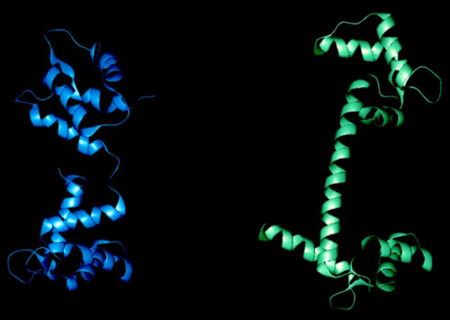
| Here you can see what happens to the protein when all four of the EF-Hands bind Ca2+ ions. The whole structure changes from the one in blue to the much tighter version in green. The central linker is held rigid in the Ca2+ form but it falls apart in the apo form. |